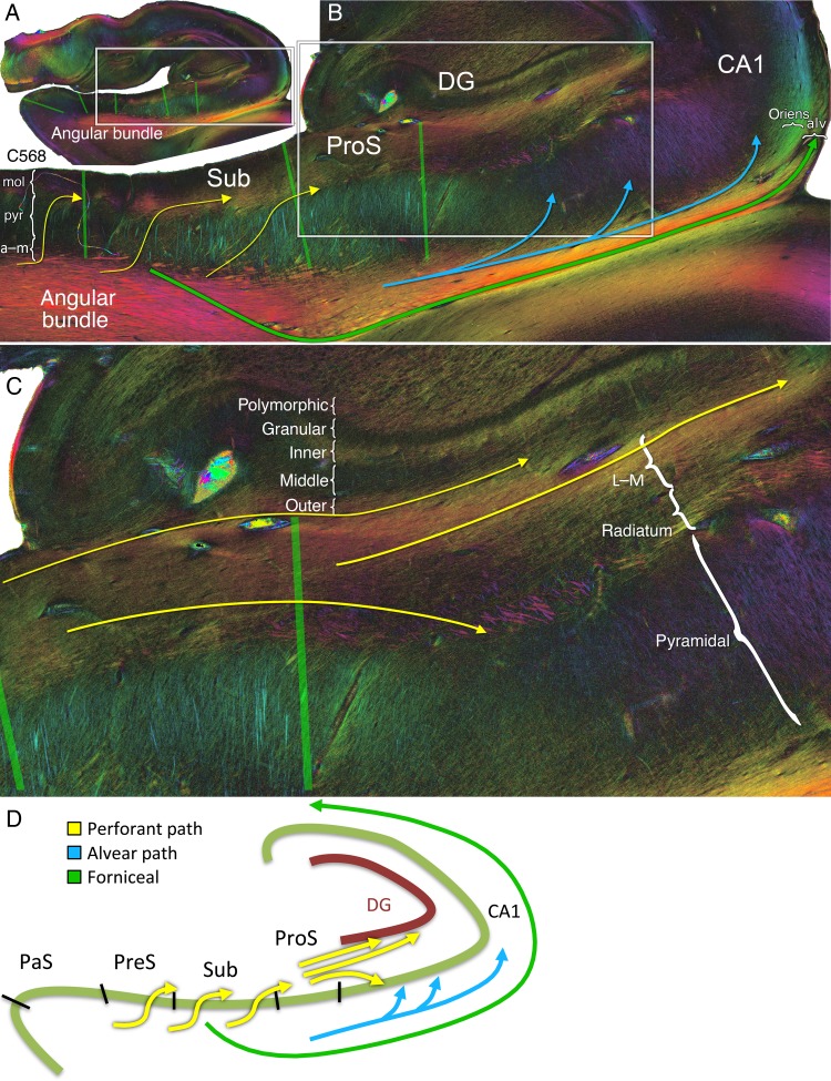
Figure 8.
Perforant path system in-plane. (A) Angular bundle fibers projecting to the more posterior portion of the hippocampal head. (B) Perforant (yellow), alvear (light blue), and forniceal (green) pathways, shown at a higher magnification (see box in A). Yellow arrows: Sigmoidal perforant pathway fibers of varying obliquities can be seen extending from the angular bundle, through the subiculum, and into the deeper part of the molecular layer of the subiculum. Light blue arrows: Deeper angular bundle alvear pathway fibers extend subjacent to the prosubiculum and into the stratum oriens where they penetrate the pyramidal layer of CA1. Green arrows: output fibers emanating from the subiculum obliquely cross the fibers of the angular bundle en route to the parahippocampal gyrus and then alveus, ultimately to reach the fornix. Subicular lamination is indicated as mol, molecular; pyr, pyramidal; a–m, alvear/multiforme. CA1 lamination is indicated as oriens, stratum oriens; alv, alveus. (C) Higher magnification of the box shown in B depicting the dentate layers (polymorphic, granular, and inner/middle/outer thirds of the molecular layer) and CA1 lacunosum-moleculare (L-M), radiatum, and pyramidal layers. Perforant pathway bifurcation: Tangential perforant pathway fibers in the superficial molecular layer of the subiculum extend into the superficial L-M and project to the outer one-third and to a lesser degree to the middle one-third of the dentate molecular layer (yellow arrows, top). Fibers in the deeper molecular layer of the subiculum continue in a tangential manner in the radiatum and then turn inferiorly to penetrate deeper into the CA1 pyramidal cell layer (yellow curved arrow, bottom). (D) Graphical depiction of the perforant, alvear, and forniceal pathways.

