Fig. 6.

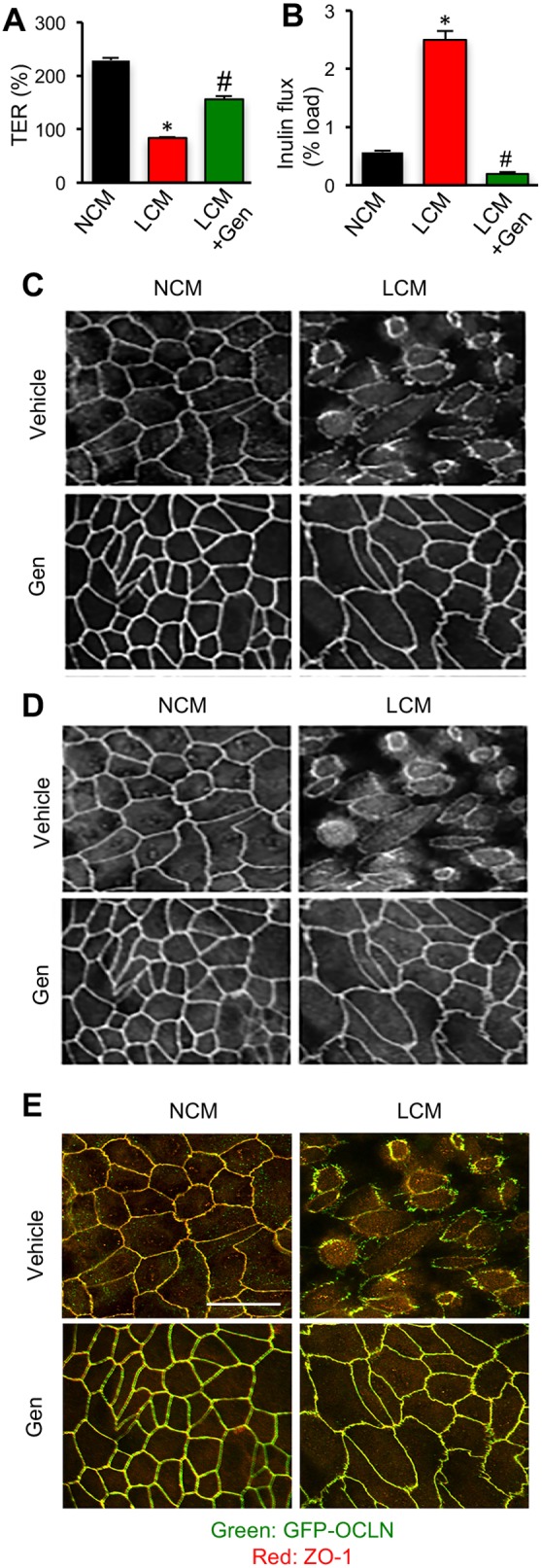
Inhibition of tyrosine kinase activity blocks Ca2+-depletion-mediated disruption of TJs and barrier function. (A,B) MDCK cell monolayers on transwell inserts were treated with 100 µM genistein (Gen) 30 min prior to LCM treatment. TER (A) and FITC-inulin flux (B) were measured after 1 h. Values are means±s.e.m. (n=6). Asterisks indicate the values that are significantly (P<0.05) different from corresponding value for OCLNWT cell monolayers and hash signs indicate the values that are significantly (P<0.05) different from that of OCLNDM cell monolayers. (C,D) Cell monolayers were co-stained for GFP-occludin (C) and ZO-1 (D). Merged images are presented in panel E. Scale bar: 50 µm.
