Fig. 5.
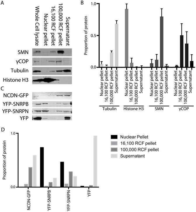
Detergent-free fractionation of SH-SY5Y cells reveals that SMN, coatomer proteins, NCDN, SNRPB and SNRPN are all enriched in the 100,000 g vesicle pellet. (A) Immunoblotting of equal protein amounts from fractionated SH-SY5Y cells reveals that SMN (top row) is highly enriched in the 100,000 g (RCF) pellet (small membrane-bound structures), with smaller amounts seen in the 16,100 g pellet (larger membrane-bound structures) and the nuclear pellet. The coatomer protein, γCOP (also known as COPG1; second row) is also enriched in the 100,000 g pellet as well as the 16,100 g pellet. Antibodies against histone H3 and tubulin confirm minimal nuclear contamination in cytoplasmic fractions, and minimal cytoplasmic contamination in the nuclear pellet, respectively. (B) Quantification of immunoblot analysis confirms that SMN is highly enriched in the 100,000 g pellet, with enrichment of γCOP also seen. Histone H3 and tubulin are highly enriched in the nucleus and cytoplasm, respectively. Quantification (mean±s.d.) of tubulin and histone H3 band density was from seven immunoblots, with values from SMN and γCOP from five and four immunoblots, respectively. (C) Immunoblotting of equal protein amounts from fractionated SH-SY5Y cells constitutively expressing NCDN–GFP, YFP–SNRPB, YFP–SNRPN or YFP alone (all detected with anti-GFP antibody) reveals that NCDN–GFP is enriched in the 100,000 g pellet, with smaller amounts seen in the 16,100 g pellet and the cytosolic supernatant. YFP–SNRPB and YFP–SNRPN are both also found in the 100,000 g pellet, in addition to the nuclear pellet and cytosolic supernatant. YFP alone is found almost exclusively in the cytosolic supernatant, with none detected in the 100,000 g or 16,100 g pellets. (D) Quantification of the band densities for the immunoblot shown in C confirms the presence of NCDN–GFP, YFP–SNRPB and YFP–SNRPN in the 100,000 g pellet, together with the restriction of YFP alone to the residual cytosolic supernatant.

