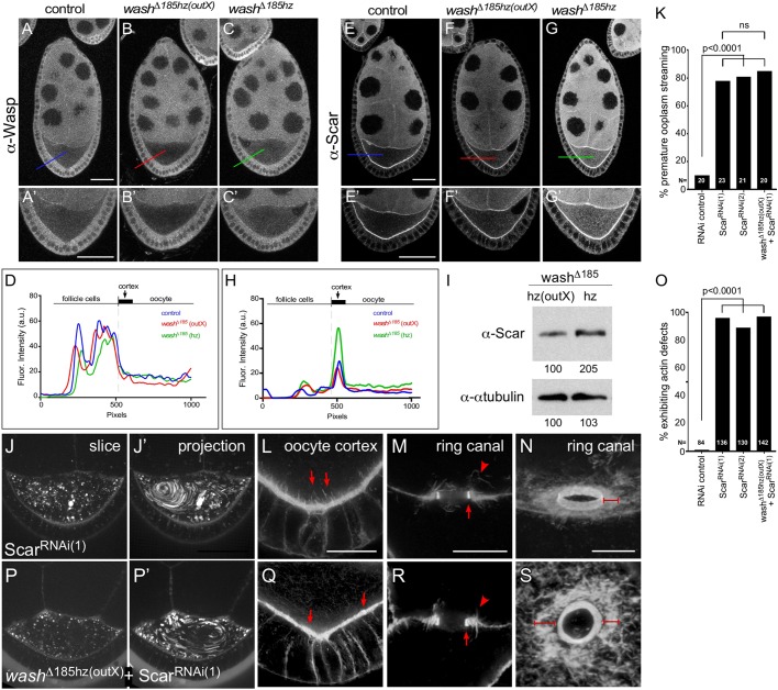
Fig. 6.
SCAR accumulates at the oocyte cortex, its loss leads to premature ooplasmic swirling and egg-chamber defects, and it is upregulated in the washΔ185 line when maintained as a homozygous stock. (A-C′) Anti-Wasp (P5E1) staining in stage 7/8 oocytes from wild-type control (A-A′), washΔ185hz(outX) (B-B′) and washΔ185hz (C-C′). (D) Line plot analysis of anti-Wasp (P5E1) staining across the cortex of control, washΔ185hz(outX) and washΔ185hz oocytes (corresponding to bars in A-C). (E-G′) Anti-SCAR (P1C1) staining in stage 7/8 oocytes from control (E,E′), washΔ185hz(outX) (F,F′) and washΔ185hz (G,G′). (E) Line plot analysis of anti-SCAR (P1C1) staining across the cortex of control, washΔ185hz(outX) and washΔ185hz oocytes (corresponding to bars in E-G). (I) Western blot analysis of ovary lysates from washΔ185hz(outX) and washΔ185hz showing increase of Scar protein levels in washΔ185hz egg chambers. Normalized quantification of each band is provided below the lane. (J,J′) Single time-point and 30 time-point projections of live time-lapse movies of stage 7 ScarRNAi(1) oocyte. (K) Quantification of the percentage of stage 7 Scar RNAi and washΔ185hz(outX)+ScarRNAi(1) oocytes exhibiting premature ooplasmic streaming (N for each genotype indicated on graph). (L) Posterior cortex of stage 7 ScarRNAi(1) egg chamber stained for F-actin (Phalloidin) in ScarRNAi(1) stage 7-9 oocyte. Note aberrantly long actin projections from cortex into oocyte (arrows). (M) Cross section of ring canal bridging the oocyte and a nurse cell in stage 7/8 ScarRNAi(1) egg chamber stained for F-actin (Phalloidin). Arrow denotes inner actin ring canal and arrowhead denotes outer actin ring. (N) Oblique view of ring canal bridging two nurse cells in stage 9 ScarRNAi(1) egg chambers stained for F-actin. Note disorganized outer actin ring (bracket). (P,P′) Single time-point and 30 time-point projections of live time-lapse movies of stage 7 washΔ185hz(outX)+ScarRNAi(1) oocyte. (Q) Posterior cortex of stage 7 washΔ185hz(outX)+ScarRNAi(1) egg chamber stained for F-actin (Phalloidin). Note uneven actin projections from cortex into oocyte (arrows). (R) Cross section of ring canal bridging the oocyte and a nurse cell in stage 7/8 washΔ185hz(outX)+ScarRNAi(1) egg chamber stained for F-actin (Phalloidin). Arrow denotes inner actin ring canal and arrowhead denotes outer actin ring. (S) Oblique view of ring canal bridging two nurse cells in stage 9 washΔ185hz(outX)+ScarRNAi(1) egg chambers stained for F-actin. Note uneven outer actin ring (brackets). Two-tailed Fisher's exact test (K,O). Sample size (N) indicated in each figure panel; ns, not significant. Scale bars: 50 µm in A-G′,J,J′,O,O′; 25 µm in L,Q; 5 µm in M-N, R-S.

