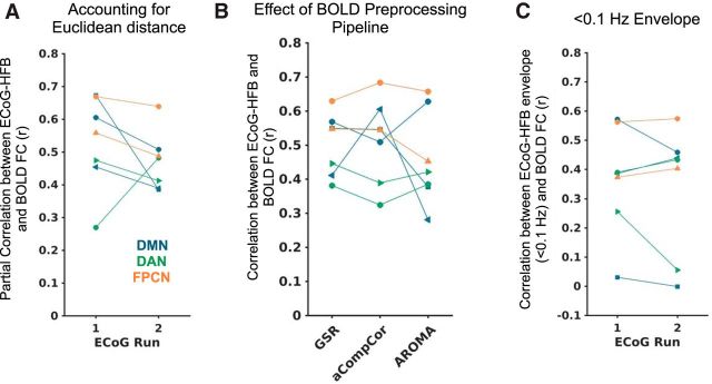
Figure 2.
Within-subject spatial correlation between BOLD and ECoG-HFB functional connectivity when A) controlling for Euclidean distance between the seed region and each target region via partial correlation (and using 0.1–1 Hz HFB envelopes), B) performing different preprocessing pipelines on the fMRI data (and using 0.1–1 Hz HFB envelopes), and C) using <0.1 Hz HFB envelopes. Each subject is indicated with a different marker shape, and each network is labeled with a different color.

