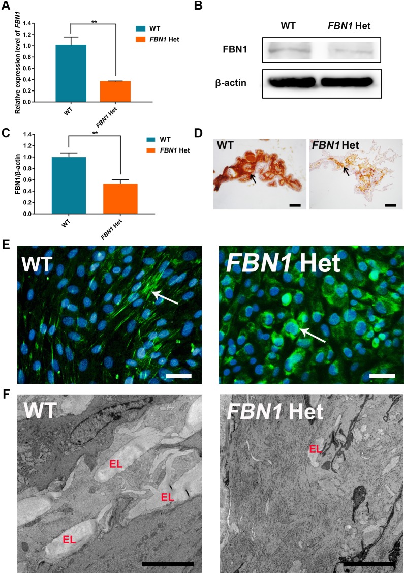
Fig. 2.
Reduced expression and secretion of fibrillin-1 in FBN1 Het rabbits. (A) qPCR results show the significantly decreased expression of FBN1 in FBN1 Het rabbits. n=5. (B) Western blot showing significantly decreased expression of fibrillin-1 protein in FBN1 Het rabbits. (C) Gray-scale analysis results show significantly decreased expression of fibrillin-1 protein in FBN1 Het rabbits. n=5. (D) IHC showed decreased expression of fibrillin-1 (arrow) in the ciliary body of FBN1 Het rabbits. (E) Immunofluorescence showed decreased secretion and assembly of microfibrils (arrow) into the extracellular matrix in FBN1 Het rabbits. (F) Transmission electron microscopy showed reduced elastin (EL) in the thoracic aorta of FBN1 Het rabbits. Data are presented as mean± s.e.m. and analyzed using Student's t-tests with GraphPad Prism software 6.0. **P<0.01. Scale bars: 50 μm.

