Fig. 3.
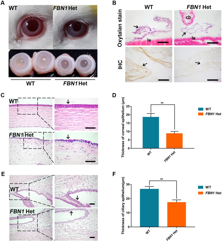
Ocular symptoms in FBN1 Het rabbits. (A) The appearance and anatomy of FBN1 Het rabbit eyes, showing the enophthalmus, eyelid abnormalities and microphthalmia. (B) Oxytalan staining and IHC show the presence of zonules (arrows) and decreased fibrillin-1 in zonules of FBN1 Het rabbits. cb, ciliary body; le, lens epithelium. (C) H&E stain showing the defect in corneal epithelium (arrows) in FBN1 Het rabbits. (D) Statistical comparison of the thickness of the corneal epithelium in FBN1 Het rabbits and WT controls. (E) H&E stain showing the defect in nonpigmented epithelium in the ciliary body (arrow) in FBN1 Het rabbits. (F) Statistical comparison of the thickness of nonpigmented epithelium in the ciliary body in FBN1 Het rabbits and WT controls. Data are presented as mean±s.e.m. and analyzed using Student's t-tests with GraphPad Prism software 6.0. **P<0.01. n=6. Scale bars: 1 μm.

