Fig. 5.
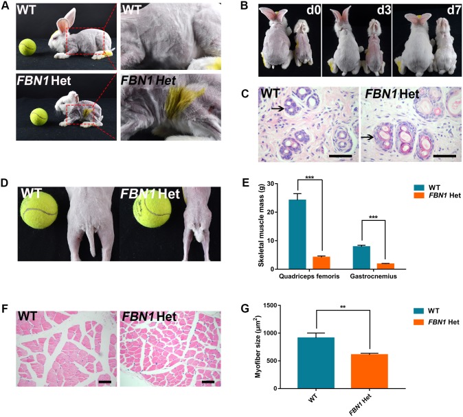
Skin symptoms and muscle wasting in FBN1 Het rabbits. (A) Photograph showing atrophic and loose skin in the FBN1 Het rabbits. (B) Photograph showing slower speed of hair re-growth in the FBN1 Het rabbits compared with WT controls (left, WT control; right, FBN1 Het rabbit). ‘d0’ represents 0 day after shaving hair; ‘d3’ represents 3 days after shaving hair; ‘d7’ represents 7 days after shaving hair. (C) H&E stain showing the degenerated hair follicles (arrow) of FBN1 Het rabbits (n=6). (D) Photograph showing the slender skeletal muscles of FBN1 Het rabbits. (E) Weight comparison results show the significantly reduced gastrocnemius and quadriceps sizes in FBN1 Het rabbits. n=6. (F) H&E staining and statistics show significantly slender myofibers in FBN1 Het rabbits. (G) Statistical analysis showing thinner myofibers in FBN1 Het rabbits compared with those in WT controls. n=30. Data are presented as mean±s.e.m. and analyzed using Student's t-tests with GraphPad Prism software 6.0. **P<0.01; *** P<0.001. Scale bars: 50 μm.

