
Fig. 2.
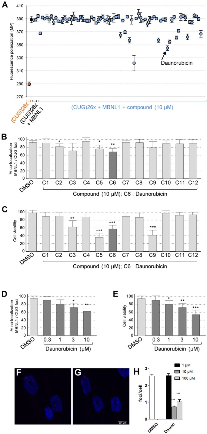
Validation screening of the 76 compounds tested at a concentration of 10 µM in DMSO to identify molecules reducing binding of MBNL1 to expanded-CUG-repeat RNA. (A) Fluorescence polarization of TRITC-labelled (CUG)26× RNA alone or in complex with GST-MBNL1-HIS recombinant protein is indicated by orange or blue dots, respectively. N=3 independent assays. (B) Percent of CUG RNA foci presenting a colocalization with endogenous MBNL1 in cultures of DM1 myoblasts upon drug treatment at 10 µM in DMSO for 24 h. N=3 independent cultures; 30 RNA foci were analyzed in each experiment. (C) Percent of living DM1 myoblasts upon drug treatment at 10 µM in DMSO for 24 h. (D) Percent of CUG RNA foci showing colocalization with endogenous MBNL1 in cultures of DM1 myoblasts upon daunorubicin treatment at 0.3, 1, 3 and 10 µM in DMSO for 24 h. N=4 independent cultures; at least 50 cells were analyzed each time. (E) Percent of living DM1 myoblasts upon daunorubicin treatment at 0.3, 1, 3 and 10 µM in DMSO for 24 h. (F-H) Foci detection in DM1 fibroblasts. Representative confocal images of FISH in DM1 fibroblasts treated with DMSO as control (F) or daunorubicin (G) showed reduced number of foci (red in F and G) with daunorubicin treatment. Nuclei were counterstained with DAPI (blue). (H) Quantification of foci confirmed a statistically significant difference at concentrations of daunorubicin higher than 1 µM. For all figure panels, error bars indicate s.e.m. Student's t-test: *P<0.05; **P<0.01; ***P<0.001.