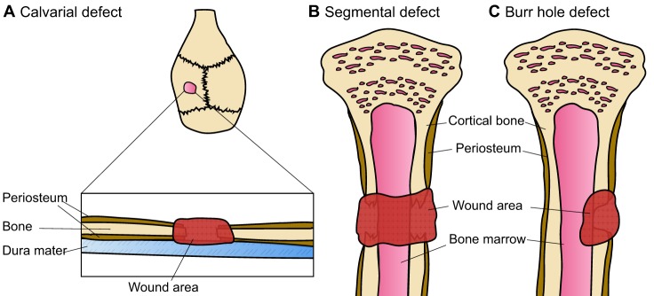
Fig. 1.
Prevalent bone defect models. (A) Calvarial defects are generally created via the introduction of a circular burr hole and the subsequent removal of the resulting bone disk. The surgery is performed in a manner so as to not damage the underlying dura mater. (B) In the segmental bone defect model, a larger and completely penetrating bone defect is generated. A segment of the bone is surgically removed, leaving a large and non-joining wound area (gap) between the bone edges. The gap is usually stabilised with a fixation device and/or filled with a tissue-engineered bone substitute to stimulate bone healing and to study bone formation. (C) In the burr hole, or partial defect model, an incomplete hole is drilled into the side of the bone to create a wounded area. The burr hole usually penetrates the cortical bone and can extend into the underlying cancellous bone or the bone marrow cavity. In this model, usually only one side of the bone is wounded.

