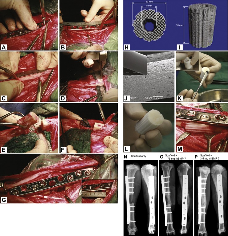
Fig. 2.
Surgery and scaffold/rhBMP-7 preparation. (A-G) Surgical generation of a segmental bone defect and implantation of a TE scaffold. To create a 3 cm segmental tibial defect, the bone was exposed and a dynamic compression plate was temporarily fixed with two screws (A). Subsequently, the screw holes were drilled, the defect middle and osteotomy lines were marked (B,C), and the bone segment was removed after osteotomy (D,E). The periosteum was removed 1 cm on the either end of the tibia defect site before the bone fragments were realigned (F) and fixed with plate and screws (G). (H-M) Top (H) and lateral (I) views of a cylindrical medical grade polycaprolactone tricalcium phosphate (mPCL-TCP) scaffold produced via fused deposition. Prior to transplantation, the scaffolds were surface treated with NaOH to render them more hydrophilic, as demonstrated in the scanning electron microscopy images prior to (J, inset) and after (J) NaOH treatment. To load the scaffolds with the recombinant human bone morphogenic protein BMP-7, the lyophilised BMP-7 was mixed with sterile saline and transferred to the inner duct of the scaffold and onto the contact interfaces between the bone and the scaffold (K,L). The BMP-7-augmented scaffolds were then implanted into the segmental tibial defects (M). (N-P) Representative X-ray images showing segmental tibial defects after 3 months of treatment with the scaffold only (N), the scaffold augmented with 1.75 mg rhBMP-7 (O) or the scaffold augmented with 3.5 mg rhBMP-7 (P), showing superior bone regeneration in the scaffolds with increasing amounts of rhBMP-7 loading due to the potent osteoinductive properties of rhBMP-7. Adapted from Cipitria et al. (2013).

