Fig. 1.

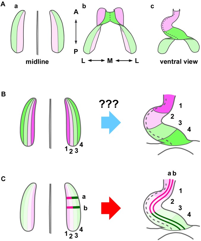
Heart fate maps. (A) Kelly et al. (2001) have revealed that the progenitors that form the anterior region (pink) of the heart tube (Ac) originally reside in the medial part (pink) of the bilateral heart primordia (Aa), which is the so-called second heart field (SHF, pink), as opposed to the first heart field (FHF, green), the lateral part of the primordia. (B) Abu-Issa and Kirby (2008) proposed that the progenitors that form the corresponding anterior-to-posterior levels of the heart tube are initially organized in medial-to-lateral sequence in each heart primordium. However, how the primordia undergo such a dramatic morphogenetic rearrangement remains unknown (question marks). (C) Our new model based on cell cluster labeling data in this study (Figs 2 and 3) showing that the heart primordia undergo dynamic tissue reshaping through CE. Areas 1 (magenta) and 2 (pink) constitute the SHF and areas 3 (light green) and 4 (green) constitute the FHF. A, anterior (cranial); P, posterior (caudal); M, medial; L, lateral.
