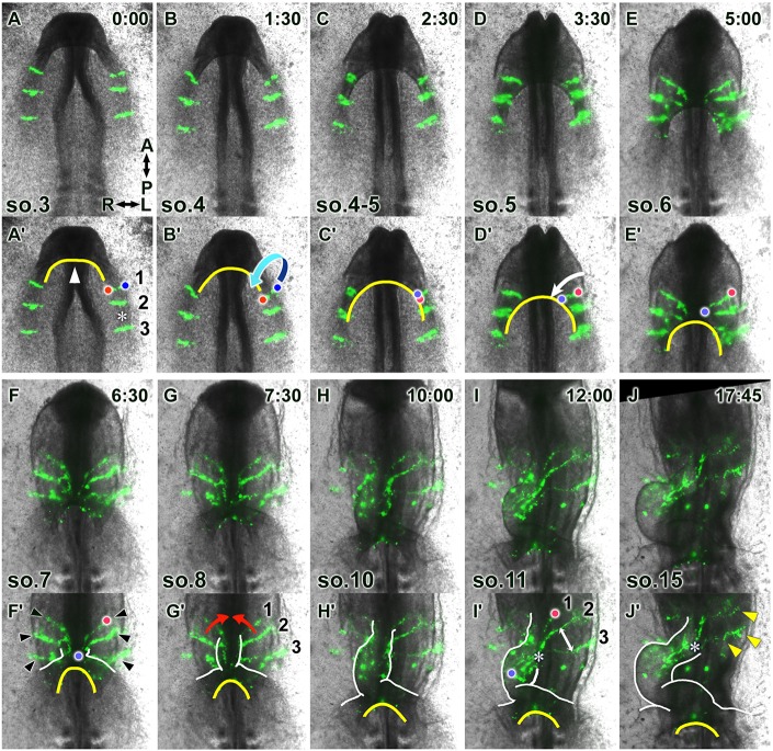
Fig. 2.
The heart primordia undergo rapid directional extension while converging perpendicularly during heart tube formation. To visualize tissue dynamics during heart tube formation, the medial parts of the bilateral heart primordia (MHP) were labeled with DiO and imaged using epifluorescence time-lapse microscopy (Movie 1). (A-J) Selected images from the movie (ventral view), with relative times after the initiation of the recording (h:min) and somite stages indicated in the upper right and bottom left corners, respectively. (A′-J′) Annotated duplicate images. Red and blue dots in A′-F′,I′ depict the original medial and lateral edges, respectively, of the labeled cell stripe. In concert with the movement of the anterior intestinal portal (AIP, yellow lines), the paired heart primordia folded ventrally, diagonally in the medial-posterior direction (arrow, B′), reorienting the original transverse cell stripes longitudinally (C′-E′). During folding, each heart primordium extended medioposteriorly (arrow, D′), along with convergence and descent of the AIP. Subsequently, the paired primordia merged medially (red arrows, G′), further reorienting the cell streams to the anteroposterior direction. White lines in F′-J′ depict the outline of the heart tube. The distance between labeled cell stripes (asterisk in A′) decreased after the heart tube formed (asterisk in I′,J′), whereas the cell stripes, which had not yet incorporated into the heart tube, remained separated (double-headed arrow in I′). At the end time-point, some MHP-derived labeled cells remained in the dorsal heart mesoderm (arrowheads in J′) in continuity with the labeled cell stripes within the anterior heart tube.

