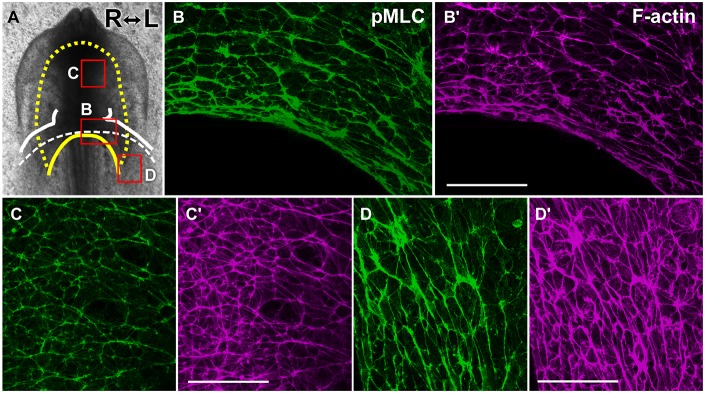
Fig. 6.
Phosphorylated-myosin is abundantly localized in a planar polarized pattern in the foregut endoderm. (A) Boxes indicate approximate areas shown in B-D′. The yellow dashed line and solid yellow line delineate the foregut and AIP, respectively. White solid lines outline the primitive heart. A white dashed line depicts the folding edge of lateral heart primordia (LHP). (B,C,D) Immunofluorescence for pMLC in normal embryos at stage 9− (6-somites, 3D projections of confocal z stacks). (B′,C′,D′) F-actin (magenta) was counterstained with fluorescent phalloidin. Phosphorylated myosin (p-myoII) was enriched in cell junctions aligned mediolaterally in the foregut (C,C′). Robust p-myoII cables were oriented circumferentially near the AIP (B,B′) and at more-posterior regions (D,D′) where the endoderm overlies the heart primordia before folding. Scale bars: 50 µm.

