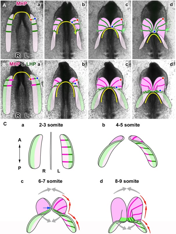
Fig. 7.
Tissue dynamics of the heart primordia during formation of the heart tube. (A) Schematic representation of heart tube formation from the medial heart primordia (MHP, pink), based on the cell labeling shown in Fig. 2. The DiO-labeled cell streams are drawn with green lines. Red and blue dots depict the original medial and lateral edges of the MHP, respectively. (B) Schematic representation of tube formation from the MHP (pink) and LHP (the lateral heart primordia; green), based on observations shown in Fig. 3. Magenta and green lines represent DiI- and DiO-labeled cell streams, respectively. Red and blue dots depict the original medial and lateral edges of the entire (MHP+LHP) heart primordium. (C) Diagrams showing the process of early heart tube formation. The paired heart primordia progressively fold, diagonally from their medial (MHP, pink) to lateral (LHP, green) sides. The first fusion of the paired primordia occurs near the boundary between the MHP and LHP (c, blue arrow), and proceeds bidirectionally (gray arrows). Whereas the MHP (pink) complete folding prior to their fusion, the folding and fusion of the LHP occur simultaneously. Both the MHP and LHP converge along the original AP axis (red arrows) as they form the heart tube while extending perpendicularly. In all drawings in A-C, the original ventral and dorsal sides of the primordia are shown in different shades of the same color to indicate how the primordia flip/fold.

