. 2018 May 10;7:e35258. doi: 10.7554/eLife.35258

Figure 2. Distinct wing disc phenotypes caused by JPS-associated tkv mutations.

(A) Human BMPR1A and Drosophila Tkv protein structures. Conserved JPS-associated mutations are indicated. (B) Scheme for the induction of tkv hetero- and homozygous mutant cells by heat shock. (C–L) wild-type and mutant forms of Tkv-mCherry (red) together with p-Mad (grey) expression in either hetero- (C–G) or homozygous (H–L) wing discs. Scale bars: 100 μm. (M) Averaged p-Mad intensity plot profiles for wild-type tkvmCherry (n = 16), tkvC90R-mCherry (n = 15) and tkvM442T-mCherry (n = 16) homozygous wing discs. 0 indicates the compartment boundary position (posterior is shaded). (N–Q) High levels of BMP activity observed in residual wild-type clones within predominantly tkvC97R-mCherry homozygous wing discs. Ectopic p-Mad expression (grey) is detected in rounded clusters of wild-type cells (green in N, P). (P and Q) show magnified images of the boxed region in (N). Scale bars: 50 μm for (N), 20 μm for (P). Anterior is oriented to the left side of all images.

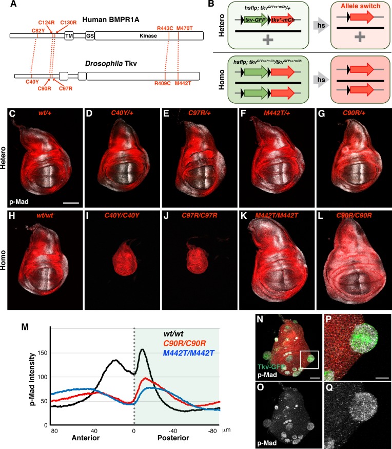
Figure 2—source data 1. BMP/DPP activation in wild-type clones within the tkvC97R homozygous background.
Wild-type clones (green) showed higher BMP/DPP activity (grey) compared to their tkvC97R homozygous neighbors (red) and frequently exhibited an outgrowth phenotype.
elife-35258-fig2-data1.docx (274.4KB, docx)
DOI: 10.7554/eLife.35258.008

Figure 2.

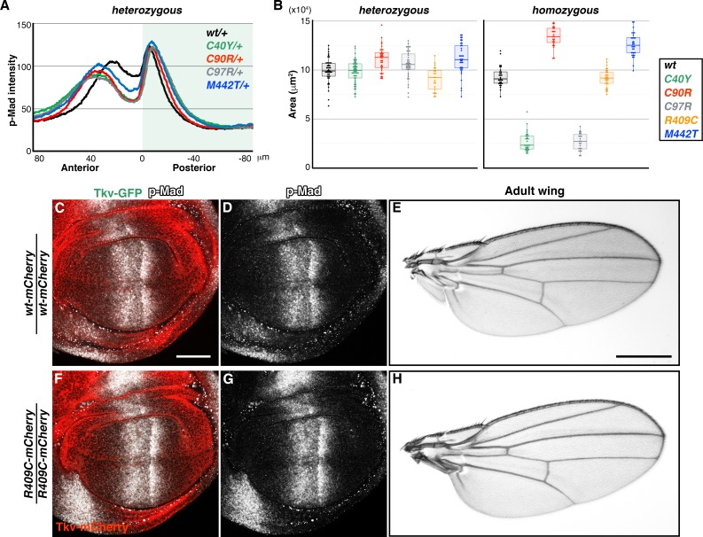
Figure 2—figure supplement 1. Phenotypes of tkv mutations.

Figure 2—figure supplement 1.

(A) Averaged p-Mad intensity profiles of wild-type tkvmCherry (n = 20), tkvC40Y-mCherry/+ (n = 24), tkvC90R-mCherry/+ (n = 24), tkvC97-mCherry/+ (n = 24), and tkvM442T-mCherry/+ (n = 16) wing discs. (B) Wing disc size comparisons. wild-type tkvmCherry (nhet = 44; nhomo = 31), tkvC40Y-mCherry (nhet = 52; nhomo = 32), tkvC90R-mCherry (nhet = 35; nhomo = 32), tkvC97R-mCherry (nhet = 42; nhomo = 23), tkvR409C-mCherry (nhet = 21; nhomo = 31), and tkvM442T-mCherry (nhet = 37; nhomo = 33). (C–H) tkvR409C homozygous animals developed normal wings. Wing discs and adult wings from wild-type tkvmCherry (C–E) and tkvR409C-mCherry (F–H) homozygous animals. Scale bars: 50 μm for (C), 0.5 mm for (E). Anterior is oriented to the left in (C), (D), (F and G), and to the top in (E and H).

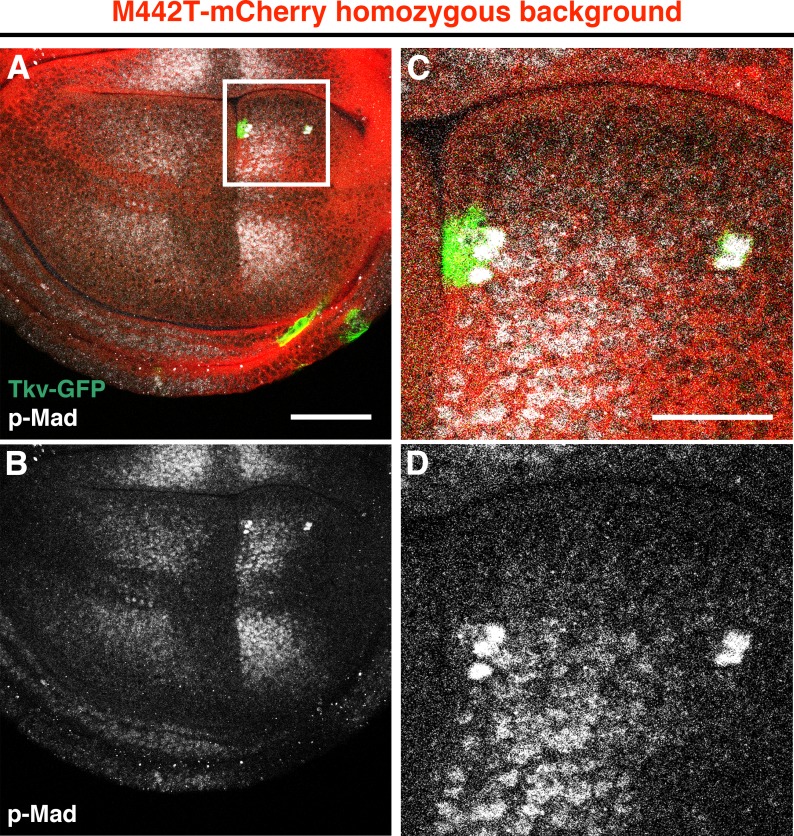
Figure 2—figure supplement 2. Strong BMP activation in cells expressing wild-type Tkv within the tkvM442T background.

Figure 2—figure supplement 2.

(A–D) Small residual wild-type Tkv-GFP clones in tkvM442T-mCherry homozygous wing discs. Tkv-GFP (green) cells in the wing pouch showed a higher p-Mad (grey) expression than tkvM442T mutant cells. Intriguingly, wild-type clones did not appear to proliferate normally in this background. Magnified images of the boxed area in (A) (C, D). Scale bars: 50 μm for (A), 20 μm for (C). Anterior is oriented to the left side of all images.