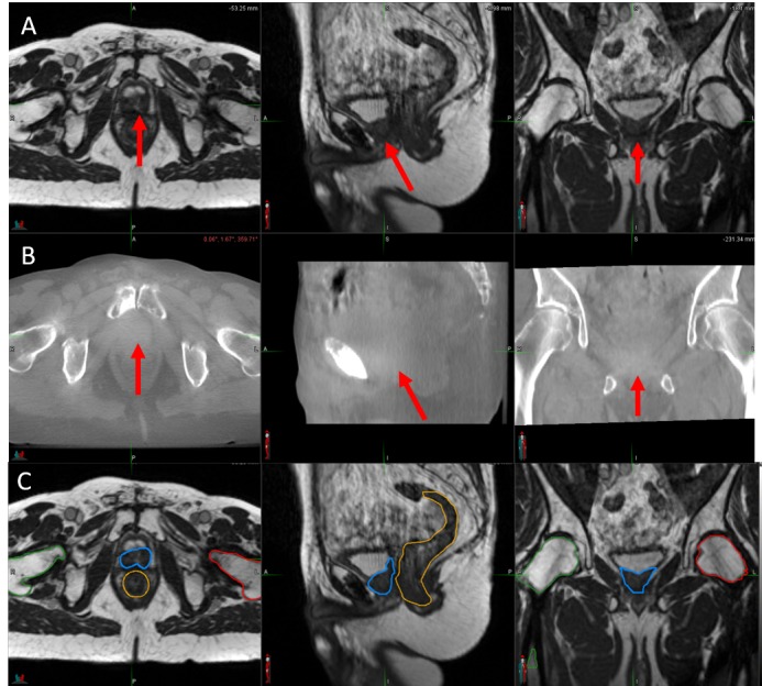
Figure 2. Image-guided Radiotherapy Images.

(A) Daily setup magnetic resonance imaging (MRI) scan obtained using the 0.35 Tesla ViewRay (ViewRay, Inc., Cleveland, OH, USA) system MRI, with a balanced steady state free precession (bSSFP) sequence. (B) Cone beam computed tomography imaging obtained using the onboard imager for the NovalisTx (BrainLAB AG, Feldkirchen, Germany) linear accelerator. In both panels (A) and (B), the red arrows indicate the location of the bulky recurrence. (C) Simulation bSSFP sequence MRI obtained on the ViewRay system, with the gross recurrent lesion depicted in blue, the rectum in orange, and the right and left femoral heads in green and red, respectively.
