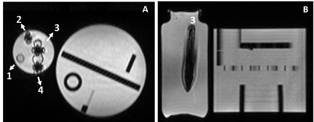
Figure 3. Grape-nuts in phantom.
Coronal (A) and axial (B) MRIs of Grape-nuts cereal (mixed with water or saliva) phantoms placed inside a 1 L bottle filled with sodium polyacrylate hydrogel. MRIs were acquired at 0.35 T using the 3D TrueFISP sequence. The 1 L bottle contained four 15 mL phantom tubes filled with: (1) water (control); (2) a small sample of unchewed Grape-nuts with water; 3) chewed Grape-nuts with saliva and water; and 4) unchewed Grape-nuts with water. The ACR phantom was placed next to the phantoms to load the body coil. The signal inside Phantom 3 is saturated (right) and the size of the phantom tube appears larger than its true dimension. Magnetic field lines appear as null bands in the coronal image for the three tubes containing Grape-nuts.
MRI: magnetic resonance imaging; ACR: American College of Radiology

