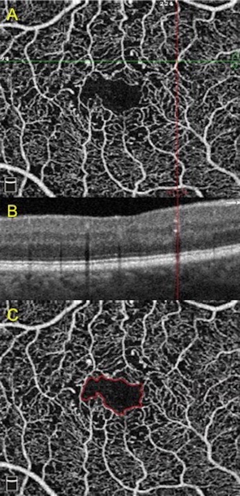
Figure 3. Examples of changes to foveal avascular zone and presence of microaneurysms.
A) Microaneurysm at intersection of horizontal and vertical lines.
B) Corresponding microaneurysm as hyperreflective spot in the inner retina marked by the vertical line.
C) Delineation of the foveal avascular zone in solid line.

