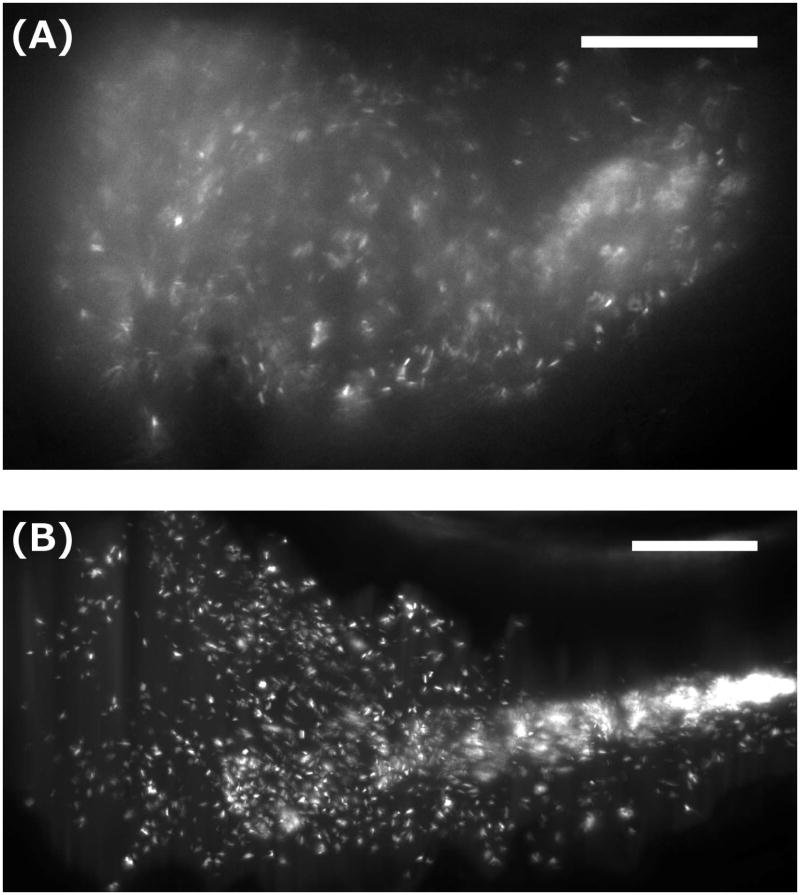
Figure 2.
Light sheet fluorescence microscopy images of bacteria in the larval zebrafish gut. (A) A single optical plane showing a GFP-expressing, commensal Plesiomonas species in the anterior intestinal bulb. Motile individuals (see Supplemental Movie 1) and sparse, likely mucus-rich aggregates are evident. (B) A maximum intensity projection of a three-dimensional image of a commensal Aeromonas species, as in Ref. [Wiles, Jemielita, et al. 2016]. Discrete individuals and a dense, midgut-localized aggregate are evident. (See Supplemental Movie 2 for a rotating 3D representation of the dataset.) Bar = 50 microns, in both panels.

