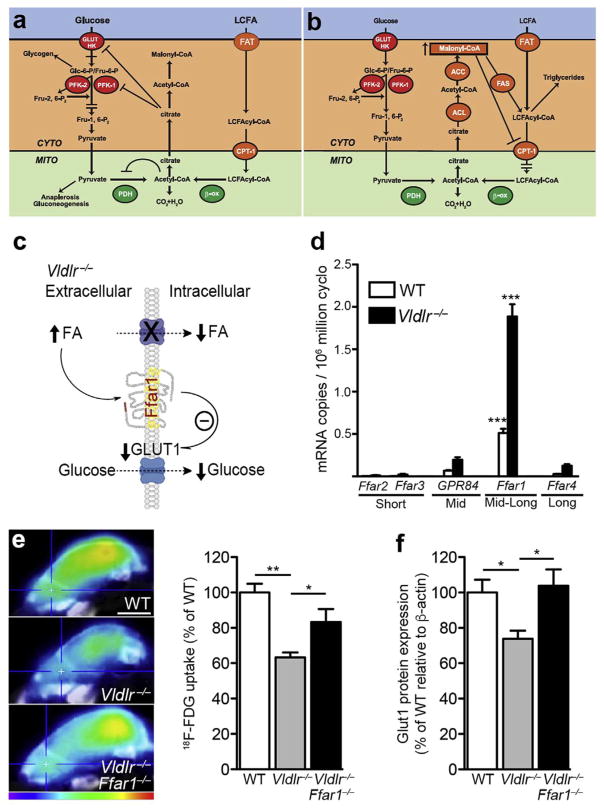
Fig. 5. Choosing between lipids and glucose as fuel: the Randle cycle and fatty acid receptors.
(a) Randle cycle: inhibition of glucose utilization by fatty acid oxidation. Accumulation of acetyl-CoA and NADH from FA oxidation inhibits pyruvate dehydrogenase (PDH), whereas cytosolic citrate regulates 6-phosphofructo-1-kinase (PFK) activity. Glucose uptake regulation is not fully explained by the Randle cycle. (b) Randle cycle: inhibition of fatty acid oxidation by glucose. Malonyl-CoA, which is produced by ACC when glucose is abundant, governs the expression of CPT1, hence regulating the entry of long-chain FA into the mitochondria. This effect re-routes fatty acids toward esterification and storage. CYTO: cytosol; MITO: mitochondria; GLUT: glucose transporter; HK: hexokinase; Glc-6-P: glucose 6-phosphate; Fru-6-P: fructose 6-phosphate; CPTI: carnitine palmitoyltransferase I; β-ox: β-oxidation, ACC: Acetyl-CoA carboxylase, ACL: ATP-citrate lyase; FAS, fatty acid synthase. (c) Elevated circulating fatty acid levels, as seen in Vldlr−/− retina, activate fatty acid receptors (such as Ffar1) that suppress GLUT1 expression and glucose uptake when lipids are abundant. (d) FA sensing receptors are expressed in WT and Vldlr−/−retinas (qRT-PCR). ONL: outer nuclear layer, INL: inner nuclear layer, GCL: ganglion cell layer. n = 3 animal retinas. (g) Glucose uptake (18F-FDG, scale: 4 mm) and Glut1 protein expression of WT and Vldlr−/− mice compared to littermate Vldlr−/−/Ffar1−/− mice (P16). Figure modified, with permission, from (Hue and Taegtmeyer, 2009; Joyal et al., 2016).

