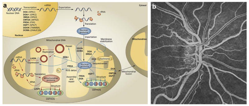
Fig. 6. Mitochondrial disorders and vascular adaptation.
(a) Cellular homeostasis is under the dual control of nuclear (blue) and mitochondrial genomes. Mitochondrial diseases (bold text), many of which impact vision, arise from mutations in genes (italic text) from either genome. Mitochondrial DNA encodes 13 structural subunits of the electron transport chain (complexes I, III, IV, and V) and the RNA needed for gene translation. All other mitochondrial proteins are encoded by the nuclear genome. Abbreviations: CMT2A, Charcot-Marie-Tooth disease, type 2A; DCMA, dilated cardiomyopathy with ataxia; DDON, deafness, dystonia and optic neuropathy; DOA, dominant optic atrophy; DOA+, dominant optic atrophy-plus syndrome; FRDA, Friedreich ataxia; HSP7, hereditary spastic paraplegia, type 7; LHON, Leber hereditary optic neuropathy; OXPHOS, oxidative phosphorylation; ROS detox, reactive oxygen species detoxification; rRNA, ribosomal RNA; tRNA, transfer RNA; 3MGA, 3-methylglutaconic aciduria, type III. (b) Fluorescein angiography of fundus during the acute stage of LHON, showing tortuous vessels and telangiectasias. Energy metabolism has a direct impact on the retinal vascular phenotype. Figure modified, with permission, from (Newman, 2012).

