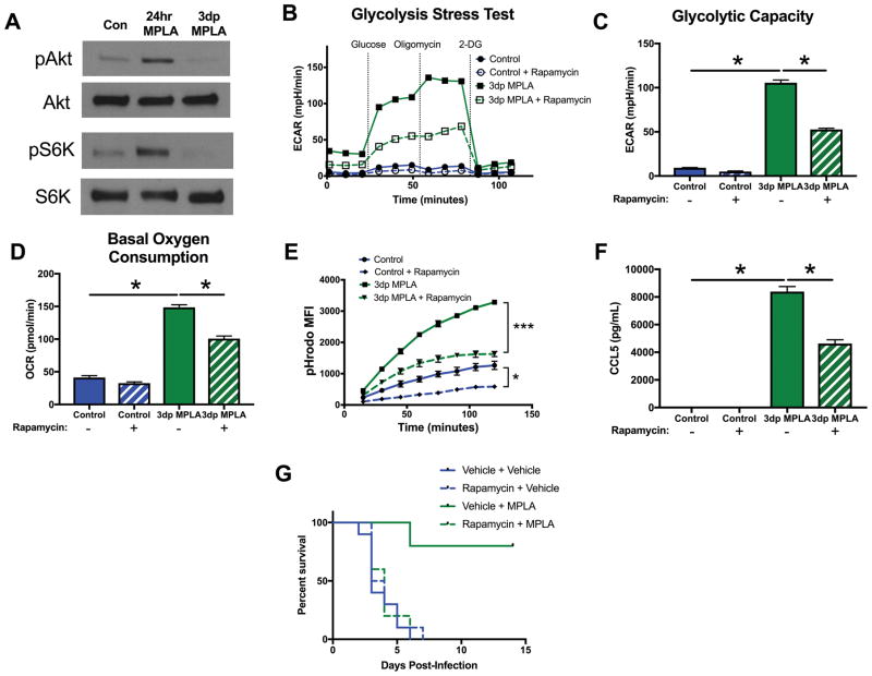
Figure 7. mTOR-initiated metabolic reprogramming is required for MPLA-induced resistance to infection.
A) Western blot of phosphorylated Akt (pAkt), total Akt (Akt), phosphorylated S6K (pS6K) and total S6K (S6K). Blots representative of 3 repeated experiments. BMDMs were exposed to 100nM rapamycin 1 hour prior to MPLA priming and throughout the 24 hour priming period. Rapamycin was washed out after priming along with MPLA. B) Glycolysis stress test of 3dp macrophages with and without rapamycin during priming. C) Glycolytic capacity of 3dp macrophages with and without rapamycin during priming. D) Basal OCR of 3dp macrophages with and without rapamycin during priming. E) Phagocytosis over time of pHrodo-tagged S. aureus bioparticles by 3dp macrophages with and without rapamycin during priming. F) CCL5 (RANTES) secretion of 3dp macrophages with and without rapamycin during priming after 6 hours of incubation in media. G) Mice were administered 3mg/kg intraperitoneal rapamycin 3 hours prior to the first administration of intravenous MPLA. Kaplan Meier survival plot over 14 days following S. aureus infection (n=10 mice/group). All experiments replicated at least twice. Data shown as mean +/− SEM. *, p< .05 as determined by ANOVA with Tukey’s post-hoc multiple comparison analysis.

