
Figure 6. DR6 deficiency does not protect RGCs from somal or axonal degeneration after axonal injury.
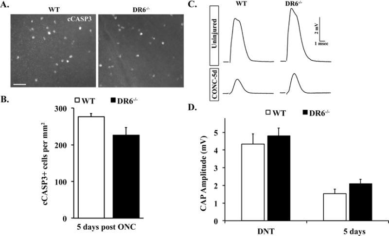
A. Representative images from WT and DR6−/− flat-mounted retinas stained with anti-cleaved caspase 3 (cCASP3) 5 days after ONC. B. Cell counts of the number of cCASP3 positive cells 5 days after crush (n ≥ 5 for each genotype) showed no significant difference between WT and DR6−/− mice (P>0.05). C. Representative traces of the compound action potential (CAP) recorded from WT and DR6−/− optic nerves 5 days after crush. D. The CAP amplitude was significantly reduced in both WT and DR6−/− optic nerves 5 days after crush. However, the reduction of CAP amplitude observed in DR6 deficient mice was similar to that observed in control mice (P>0.05; (n ≥ 5 for each genotype and condition), suggesting DR6 is not an important component of the axonal degeneration pathway in adult RGCs. Scale bar: A, 50 m; error bars, SEM).