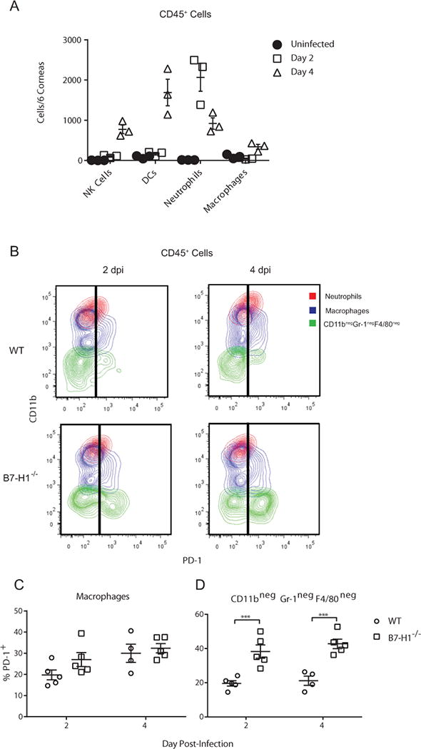
Figure 4. Expression of PD-1 by infiltrating immune cells.

(A) Corneas of wild type mice were not infected or infected with HSV-1 KOS. At 2 and 4 dpi corneas were excised, single cell suspensions of pools of 6 corneas were stained for CD45 (leukocytes), NK1.1 (NK cells), CD11c (DC), Gr-1 (neutrophils, inflammatory monocytes), F4/80 (macrophages), and for PD-1. A defined number of fluorescent beads were added to each cell sample to determine absolute cell numbers. Samples were gated on CD45 and the number of each leukocyte subpopulation in each pool of 6 corneas was determined by flow cytometry. (B-D) Corneas of wild type and B7-H1−/− mice were infected with HSV-1 KOS. At 2 and 4 dpi corneas were excised, single cell suspensions were prepared, stained for CD11b, Gr-1, F4/80, and PD-1 and analyzed by flow cytometry(B) Representative flow plots gated on CD45 cells illustrate levels of PD-1 expression on neutrophils (large, granular CD11bhigh Gr-1high F4/80−), macrophages (CD11b+, F4/80int – low), and non-myeloid cells (CD11b− Gr-1−, F4/80). Little or no PD-1 expression was detected on neutrophils. Scatter plot shows the frequency of PD-1+ cells within the (C) macrophage gate and (D) non-myeloid cell gate. The significance of differences between genotypes at individual days in C & D was assessed by a two-way ANOVA with Sidak’s post-tests (***p<0.001).
