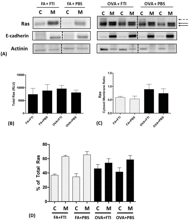
Figure 2. Subcellular Localization of Ras in Air- and Allergen-Exposed Mice and the Effects of FTI-277 Treatment on Ras Subcellular Translocation.
BALB/c mice were sensitized to ovalbumin (OVA) and then challenged with 1% OVA aerosol or filtered air six times over a 2-week period. Mice were injected daily with FTI-277 (20 mg/kg/day, i.p.) before each OVA aerosol exposure. Whole lung homogenates were processed into cytosolic [C] and membrane [M] subcellular fractions, and Ras protein expression in these fractions was assessed by Western blot. Ras semi-quantitative values were normalized using E-cadherin and actinin for membrane and cytosolic fractions, respectively.
(A) In mice exposed to FA, Ras resides predominantly in the [M] fraction. With OVA exposure, Ras becomes nearly evenly distributed between the [C] and [M] fractions. These data indicated that under basal non-inflamed conditions, Ras is farnesylated and membrane-bound. All [C] bands represent unfarnesylated Ras including the faint band above the lowest one (dashed black arrow), and the [M] bands represent farnesylated membrane-anchored Ras (solid black arrows).
The dashed black vertical lines represent the space where images of bands were joined from a single blot to show the representative bands shown here. All relevant bands (and blots), including the representative bands above, were used to generate and analyze the data shown in panels B–D.
(B) Total Ras protein expression did not differ with FTI-277 treatment for both the FA and OVA groups (p=NS by 1-way ANOVA).
(C) Plotted as cytosol-to-membrane ratio, treatment with FTI-277 did not affect Ras translocation in both FA and OVA groups (p=NS by 1-way ANOVA).
(D) Plotted as % of total Ras, treatment with FTI-277 showed no statistically significant changes in [C]- or [M]-associated Ras in both FA and OVA groups (p=NS by 1-way ANOVA). Similarly, there were no significant changes in [C] or [M] subcellular fractions in PBS controls for both FA and OVA groups (p=NS by 1-way ANOVA).

