
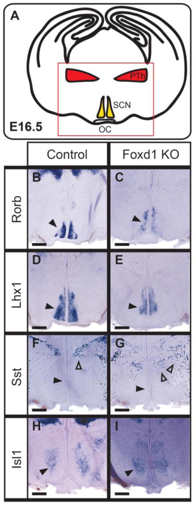
Figure 3. Progenitor proliferation, but not apoptosis, is affected by loss of Foxd1.
A–B: Immunohistochemistry staining against EdU (red) and DAPI (blue) on E12.5 heterozygote control and Foxd1 mutant coronal sections after a 2hr EdU pulse. Red scroll bars indicate the counted portion of the section containing the prethalamus and hypothalamus. C: Quantification of EdU-positive cells relative to area (μm2) in control versus Foxd1 mutant (n=3). D–E: There was no obvious change in the number of TUNEL-positive cells in Foxd1 mutants.