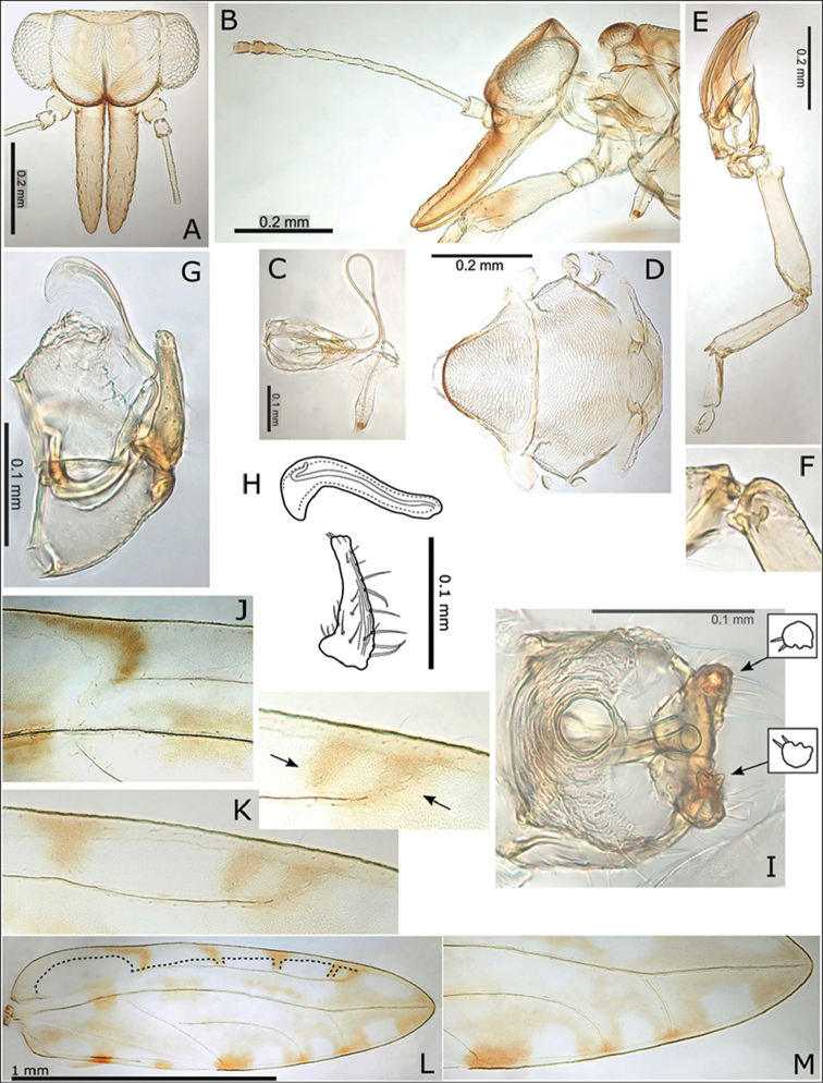
Figure 11.
Swezeyana magnaccai sp. n. A head B head and antenna (lateral view) C proboscis D dorsum of thorax E hind leg F base of hind tibia G male terminalia H aedeagus and paramere I male terminalia (dorsal view), inset details of paramere apices J fore wing detail of termination of vein R at base of pseudopterostigma K fore wing detail of incomplete termination of vein Rs at wing margin (inset incomplete veins indicated) L fore wing, with interior edge of ventral margin outlined M fore wing detail of unpigmented membrane surrounding marginal radular spine clusters.

