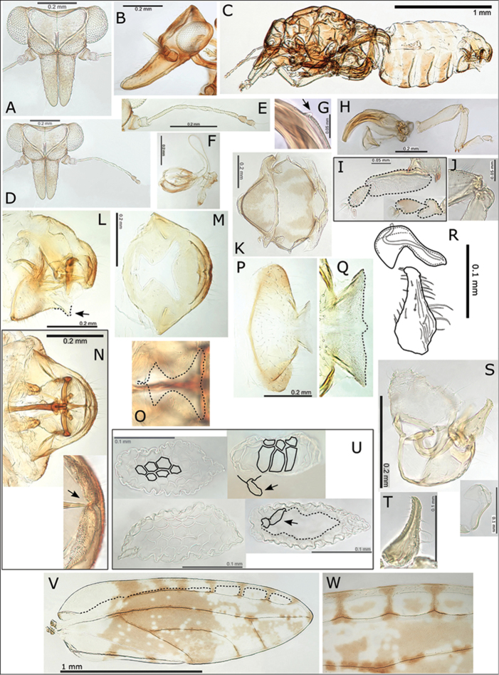
Figure 8.
Swezeyana rubra sp. n. A head B head (lateral view) C female D head and antenna E antenna F proboscis G reduced meracanthus (indicated) H hind leg I metatarsi (outlined), inset comparative size of mesotarsi (outlined) J base of hind tibia K dorsum of thorax L female terminalia (subgenital beak indicated, apex outlined) M female proctiger (dorsal view) N female terminalia (dorsal view), inset apex of proctiger (ventral view, cleft indicated) O detail of anal ring (outlined) P female subgenital plate (ventral view) Q detail of posterior apex of female subgenital plate with beak and membrane (outlined) R aedeagus and paramere S male terminalia, inset aedeagus T paramere (posterior view) U eggs (semi-hexagonal sculpturing, pedicel and unsculptured underside outlined and indicated) V fore wing, with interior edge of ventral margin outlined W fore wing detail of pigmented cross veins.

