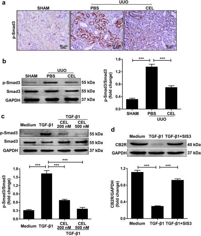
Fig. 6. Involvement of Smad3 signaling pathway in celastrol-mediated upregulation of CB2R expression.
a, b Mice received celastrol (1 mg/kg) daily for 7 days from the day of surgery. The vehicle-treated mice were treated with an equal volume of PBS. Sham group was the control for UUO mice. Kidney tissues were collected 7 days after UUO. Representative results of immunohistochemistry and western blot of p-Smad3 expression in the obstructed kidney tissue. Quantitative data were presented. c Serum-starved HK-2 cells were pretreated with or without celastrol (500 nM) for 1 h and then stimulated with TGF-β1 (10 ng/ml) for 1 h. Western blot analyses of Smad3 phosphorylation in HK-2 cells and quantitative data were presented. d Serum-starved HK-2 cells were pretreated with or without SIS3 (10 ng/ml) for 1 h and then stimulated with TGF-β1 (10 ng/ml) for 24 h. Western blot analyses of CB2R protein in HK-2 cells and quantitative data were presented. The medium group was used as the control for TGF-β1 treatment. Results are representative of three replicate experiments. All values are represented as mean ± SEM. n = 5/group. Scale bar = 50 μm. ***P < 0.001

