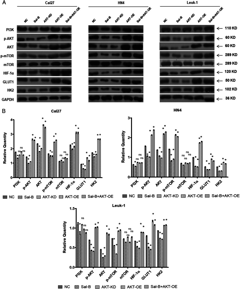
Fig. 7. Sal-B inhibited PI3K/Akt/HIF-1α signaling and glycolysis in vitro.
Cal27, HN4, and Leuk1 cells were treated with varying conditions, including (1) without treatment (NC), (2) 250 μM of Sal-B, (3) knockdown of AKT expression using AKT shRNA lentivirus (AKT-KD), (4) overexpression of AKT using transfection of AKT lentivirus (AKT-OE), and (5) the combined treatment of Sal-B and AKT-OE. a The quantitative analysis of the protein level of PI3K, p-AKT, AKT, p-mTOR, mTOR, HIF-1α, GLUT1, and HK2 in three types of cells (Cal27, HN4, and Leuk1) collected at 48 h post treatments were detected by western blotting. b Quantitive analysis from (a). P < 0.05 was considered statistically significant, ns, P > 0.05, *P < 0.05, **P < 0.01, ***P < 0.001 vs. control group

