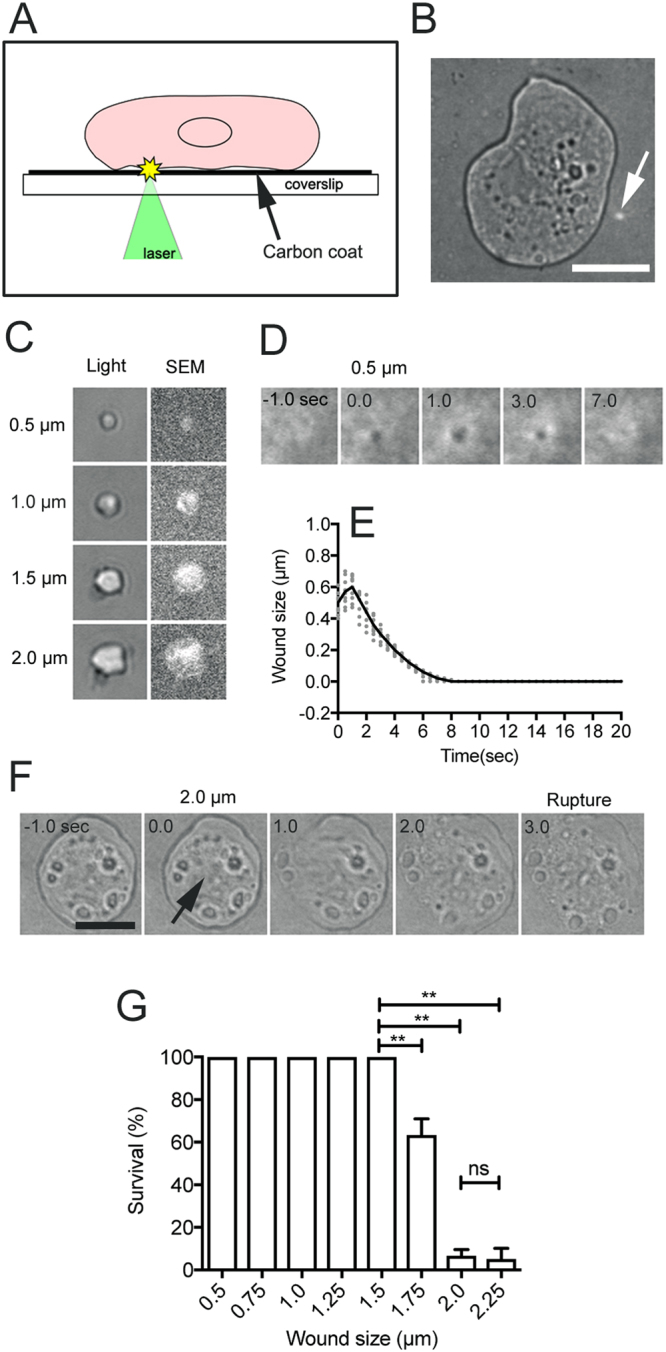
Figure 1.

A new application of laserporation to wound the cell membrane. (A) After cells were placed on a carbon-coated coverslip, a laser beam was focused on a small local spot beneath a single cell under a TIRF microscope. The absorbed energy of the laser beam by the carbon created a small pore in the cell membrane that was attached to the carbon coat. (B) When the laser beam was focused on the surface of coated coverslip, the carbon coat was peeled off, appearing as a small white spot (arrow). (C) White spots of different sizes (0.5, 1.0, 1.5, and 2.0 µm) on the carbon coat observed by light and scanning electron microscopy. (D) When the cell expressing GFP-cAR1 was wounded by laserporation, a black spot appeared in the cell membrane. The black spot transiently expanded, then shrank, and finally closed. (E) Time course of the diameter of the black spot after the laserporation. The solid line is an averaged line of 10 experiments (gray dots). (F) A typical sequence of bright field images of cell rupture with a 2 µm wound (arrow). (G) Survival rate of cells with different laser spot sizes. The number of cells that ruptured within 5 min after laserporation was counted. Data are presented as mean ± SD (n = 20, 3 experiments, each). **P ≤ 0.0001; ns, not significant, P > 0.05. Bars, 10 µm.
