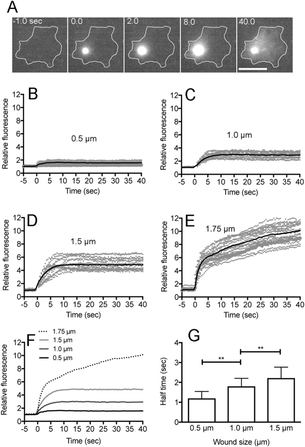
Figure 2.
Influx of fluorescent dye from the wound pore. Wound experiments were performed in BSS including propidium iodide (PI). (A) A typical sequence of fluorescence images of PI influx after laserporation. The wound laser beam was applied at 0 time and the duration was set at 8 msec. Note that the fluorescence began to increase at the wound site and spread over the cytoplasm. (B–E) Time courses of PI influx with different wound sizes (0.5, 1.0, 1.5, and 1.75 µm, n = 27, each). (F) A comparison of time courses of PI influx in 4 different wound sizes (0.5, 1.0, 1.5, and 1.75 µm). (G) Half time of closing with different wound sizes. The half time was examined by curve fitting as described in Methods section. The half time was 1.19 ± 0.34 sec with a 0.5 µm wound, 1.80 ± 0.40 sec with a 1.0 µm wound, and 2.22 ± 0.55 sec with a 1.5 µm wound after the laserporation, respectively (n = 27, each). Data are presented as mean ± SD. **P ≤ 0.0001. Bar, 10 µm.

