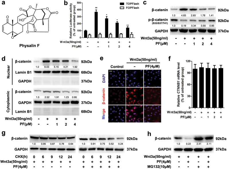
Fig. 1. PF inhibited the Wnt/β-catenin signaling.
a The chemical structure of Physalin F (PF). b Luciferase reporter activity was measured in HEK293T cells. After 24 h of transfection, HEK293T cells were treated with recombinant Wnt3a in the presence of PF at the indicated concentrations for 16 h, then followed by a luciferase assay. Data represent the means ± SD (n = 3), *P < 0.05, **P < 0.01, compared with recombinant Wnt3a group, ##P < 0.01, compared with untreated group. c HEK293T cells were treated with indicated concentrations of PF in the presence of Wnt3a for 16 h, then the β-catenin and p-β-catenin were detected by immunoblotting. d HEK293T cells were treated with indicated concentrations of PF in the presence of Wnt3a for 16 h, then the cytoplasmic and nuclear fractions lysates were analysed by immunoblotting. e HEK293T cells were treated with 4 μM PF and Wnt3a for 16 h, then the intracellular expression of β-catenin was examined by immunofluorescence, scale bars = 20 μm. f HEK293T cells were treated with indicated concentrations of PF and Wnt3a for 16 h, and the mRNA level of CTNNB1 was measured by quantitative RT-PCR. Data represent the means ± SD (n = 3). g HEK293T cells were co-treated with or without 4 μM PF and CHX (50 μg/ml) in the presence of Wnt3a for the indicated time intervals, and the expression of β-catenin was examined by immunoblotting. h HEK293T cells were treated with 4 μM PF for 12 h, then with or without 10 μM MG132 for an additional 8 h before immunoblotting

