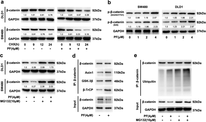
Fig. 5. PF promoted the degradation of β-catenin in CRC cells.
a SW480 and DLD1 cells were incubated with CHX (50 μg/ml) in the presence of PF or DMSO for the indicated time intervals, and the expression of β-catenin was examined by immunoblotting. b SW480 and DLD1 cells were treated with indicated concentrations of PF for 24 h, and the intracellular expression of p-β-catenin was analysed by western blot. c SW480 and DLD1 cells were incubated with 4 μM PF for 16 h, then with or without 10 μM MG132 for additional 8 h before immunoblotting. d Lysates from SW480 cells after treatment with PF (4 μM) was immunoprecipitated with β-catenin antibody, input and immunoprecipitated fractions were analysed by immunoblotting with the indicated antibodies. e SW480 cells were incubated with 4 μM PF for 16 h, then with or without 10 μM MG132 for an additional 8 h. The cell lysates were subjected to immunoprecipitation using a β-catenin antibody, and coprecipitating endogenous proteins were detected by western blot with the indicated antibodies

