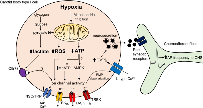
FIGURE 1.
Carotid body mitochondrial signaling mechanisms activated during hypoxia. Hypoxia-induced mitochondrial inhibition is proposed to increase lactate generation (Chang et al., 2015), augment mitochondrial complex I reactive oxygen species (ROS) production (Fernandez-Aguera et al., 2015) or reduce mitochondrial ATP synthesis (Buckler and Turner, 2013). These changes are proposed to directly or indirectly (e.g., via Olf78 receptor activation, reduced MgATP concentration or stimulation of AMP-activated protein kinase; AMPK) modify ion channel function leading to resting membrane potential (RMP) depolarization. This causes opening of L-type Ca2+ channels, neurosecretion and an increase in discharge frequency of the adjacent chemoafferent fibers. TRP, transient receptor potential channel; NSC, non-selective cation channel; BKCa, large conductance Ca2+-activated K+ channel; TASK, TWIK-related acid-sensitive K+ channel; TREK, TWIK-related K+ channel; AP, action potential.

