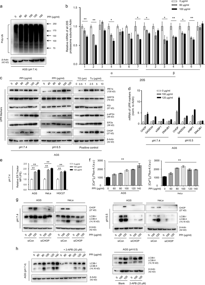
Fig. 5. PPI reduced proteasome expression and induced ER stress in cancer cells.
a AGS cells were treated with indicated concentrations of PPI for 48 h in pH 7.4 condition, followed by measuring proteasome function via western blot analysis using specific antibody to ubiquitin. b The mRNA expression level of the 20S proteasome subunits were measured after PPI treatment for 48 h in pH 7.4 condition. Data were presented as mean ± SD (*p < 0.05, **p < 0.01, for each gene using one-way ANOVA with Dunnett’s multiple comparisons test). c Western blot analysis of ER stress-related proteins after PPI treatment for 48 h in both pH 7.4 and pH 6.5 conditions. Cells treated with Thapsigargin (TG, 0.5 and 1 μM) or Tunicamycin (Tu, 2.5, 5 and 10 μg/ml) for 24 h served as positive controls. d qRT-PCR analysis of UPR genes after 48 h of PPI (100 and 120 μg/ml) treatment in both pH 7.4 and pH 6.5 conditions. Data were presented as mean ± SD (**p < 0.01, for each gene using one-way ANOVA with Dunnett’s multiple comparisons test). e ER-Tracker Red (500 nM) staining of PPI treated cells in pH 7.4 condition was performed. The quantification of ER Tracker fluorescence were accomplished by FACS. Data presented were representative of three independent experiments (**p < 0.01, for each cell line using one-way ANOVA with Dunnett’s multiple comparisons test). f After treated with PPI (60–140 μg/ml) for 24 h in pH 7.4 condition, cells were then incubated with 5 μM Fluo-4/AM and detected by flow cytometry. Data presented were representative of three independent experiments (**p < 0.01, identified by one-way ANOVA with Dunnett’s multiple comparisons test). g, h PPI-induced ER stress contributed to the acitivation of autophagy. AGS and HeLa cells transfected with CHOP-specific siRNA, were treated with 100 and 120 μg/ml PPI for 24 h in both pH 7.4 and pH 6.5 conditions (g). AGS cells were treated with the indicated concentrations of PPI in the absence or presence of 2-APB (20 μM) for 24 h in both pH 7.4 and pH 6.5 conditions (h). The autophagy marker LC3B-II was determined

