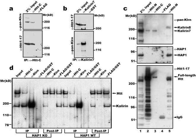
Figure 1.
Kalirin interacts with Htt in brain membranes. (a) and (b) Kalirin and Htt are mutually co-precipitated. Mouse brain membranes as described in Method were solubilized in lysis buffer containing 1% Triton X-100 and centrifuged to discard unsolubilized membranes. The supernatant was incubated with antibodies against Htt-C (aa2703-2911 of Htt)36, (a) or aa1641-1654 of kalirin-7 (b). After washes, immunoprecipitates were eluted into sample buffer and analyzed by SDS-PAGE and Western blot with indicated antibodies. Shown are blot analyses from one of four experiments for both (a) and (b). (c) and (d) Interaction between Htt and kalirin is independent of HAP1. Triton X-100 solubilized mouse brain endosomes (lanes 1, 2, and 3) or mouse embryonic stem cell total membranes (lane-5) were used for immunoprecipitation with antibodies against aa181-810 of Htt (Htt-N, lanes 2 and 5) or aa2703-2911 of Htt (Htt-C, lane-3) or the FLAG tag (lane-4). For IgG control precipitation, solubilized endosomes and ES cell membranes were mixed and incubated together with anti-FLAG antibodies (lane-4). Precipitates were washed, eluted into SDS-PAGE sample buffer and analyzed by Western blot with antibodies against pan-kalirin, HAP1, or aa1-17 of Htt. The arrowhead labeled with IgG heavy chain indicates the 55 kD band present in the complex precipitated by anti-FLAG antibodies. We noticed that the majority of Htt proteins were cleaved during experimental procedures (Input, lane-1). This was not a surprise because it is well known that NH2 Htt is prone to cleavage by proteases. This may be the reason why the antibody against Htt1-17 detected much weaker signals of full-length Htt in precipitates obtained with anti-Htt-C antibodies. (d) Triton X-100 solubilized cortical total membranes from HAP1 knockout (KO) and corresponding WT mouse embryos were incubated with antibodies specific for aa2703-2911 of Htt or kalirin7 or FLAG/GST (IgG controls). Precipitates were washed and analyzed by SDS-PAGE and Western blot with antibodies for detecting Htt and kalirin. Blots were first probed with anti-pan-kalirin to detect precipitated kalirin and then re-probed with anti-Htt1-17 to detect precipitated Htt.

