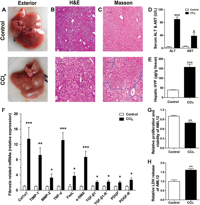
Fig. 1. Characterization of mouse model of in vivo hepatic damage induced by injection of CCl4 for 3 weeks, and of in vitro hepatocyte damage induced by CCl4 for 24 h.
a Comparison of the exterior appearance of livers between the CCl4 and control groups; (b) comparison of H&E staining of liver section between the CCl4 and control groups showing the differences of hepatic architecture; (c) comparison of Masson trichrome staining of liver section between the CCl4 and control groups showing the differences of hepatic fibrosis; the scale bars represent 10 μm. d Comparison of the contents of serum ALT and AST between the CCl4 and control groups. N = 7 for each group. e Comparison of the contents of hepatic HYP between the CCl4 and control groups. N = 7 for each group. f Alterations of expression levels of the genes related to hepatic damages in CCl4-treated mice compared with the control animals, as determined by qRT-PCR. The genes detected included Col1α1, α-SMA, TIMP-1, MMP-2, TNF-α, and TGF-β1. N = 7 for each group. g The relative cellular proliferation and viability was measured by CCK-8 assay in normal control and damaged AML12 induced by CCl4. N = 3 for each group. h Relative LDH release in normal control and CCl4-treated AML12. N = 3 for each group. All of the data were expressed as mean ± SEM. *P < 0.05, **P < 0.01, and ***P < 0.001 vs. control

