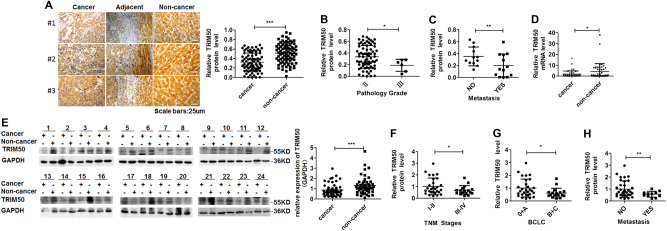
Fig. 1. TRIM50 was downregulated in HCC tissues and its expression was inversely correlated with advanced disease progression.
a Immunohistochemical staining was used to determine the location and expression of TRIM50 in HCC tissues and corresponding non-cancerous liver tissues from 79 clinical HCC patients. The intensities of the IHC staining were quantitatively analyzed by IPP6 software and statistically analyzed (right panel). b, c TRIM50 protein levels in different pathology grades (b) and different metastasis stages (c) from 79 clinical HCC patients used for IHC staining were statistically analyzed and compared. d mRNA expression of TRIM50 was determined by qRT-PCR in HCC tissues and corresponding non-cancerous liver tissues from 51 HCC patients. e Western blot analysis of protein levels of TRIM50 in the liver cancer tissues and corresponding non-cancerous liver tissues from 52 HCC patients, with GAPDH expression as internal references. The presented images are representative blots from 24 HCC patients. Band intensities of all the investigated patients were measured by Image J software and statistically analyzed (right panel). f-h Statistical analysis of TRIM50 protein level in different TNM stages (f), different BCLC stages (g), and different metastasis stages (h) from HCC patients used for western blot assay. *P < 0.05, **P < 0.01, and ***P < 0.001 for statistical analysis of the indicated groups

