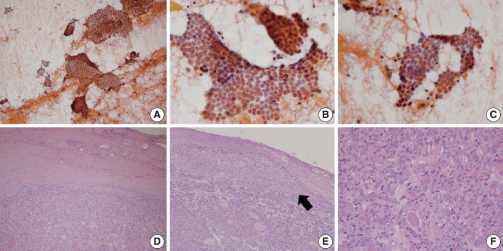
Fig. 2.

A representative example of encapsulated follicular variant of papillary thyroid carcinoma with invasion classified as papillary carcinoma in fine-needle aspiration cytology. (A) Low power examination reveals large tissue fragments and small clusters in cytologic smear. (B, C) Monolayered sheets or syncytial clusters showing nuclear elongation, overlapping, loss of polarity and grooves. (D) In the final histology of surgical excision specimen, the tumor has a thick fibrous capsule with irregular border. (E) A focus of capsular invasion (arrow) is evident. (F) Tumor cells are arranged in microfollicular or trabecular pattern, showing typical papillary thyroid carcinoma–like nuclear features.
