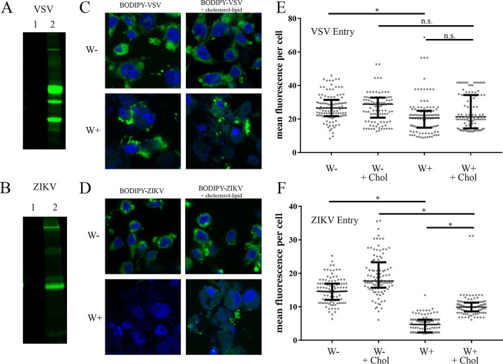
FIG 3 .
Wolbachia wStri blocks ZIKV entry. (A and B) VSV (A) and ZIKV (B) were pelleted at high speed followed by resuspension in PBS buffer. Pelleted virus was labeled with a lysine conjugate, BODIPY 650/665. Virus labeling was visualized on a 10% SDS-PAGE gel and imaged at 700 nm to confirm BODIPY incorporation in glycoprotein and envelope protein, respectively. Lane 1, unlabeled virus; lane 2, labeled virus. (C to F) Labeled virus was incubated with W+ and W− cells at an MOI of 10 for 1 h at 28°C. After fixation, cells were mounted in Prolong Gold antifade medium with DAPI. (C and D) Representative images of BODIPY-virus incorporation. BODIPY-VSV (C) or BODIPY-ZIKV (D) are shown in green. DNA stained by DAPI is blue. (E and F) Individual cells were quantified for mean BODIPY intensity per cell for VSV (E) and ZIKV (F). Cell periphery was determined by differential interference contrast imaging. Reproducibility was confirmed through independent biological replicates. Statistically significant mean fluorescence was assessed by one-way ANOVA followed by Tukey’s test for multiple comparisons. *, P < 0.05.

