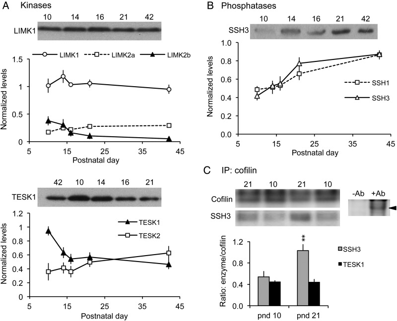
Figure 3.
Levels of the cofilin phosphatase slingshot (SSH)-3 increase during the third postnatal week. (A,B) Western blots of whole hippocampal homogenates were used to evaluate relative levels of cofilin kinases and phosphatases. For each sample, band densities were normalized to those for tubulin; group mean ± SEM values are shown for n = 4–6 animals samples per group. (A) Levels of LIMK1, LIMK2a, and TESK2 showed little change from pnd 10 to 42 (representative blots for LIMK1 and TESK1 shown). In contrast, LIMK2b levels declined from pnd 10 to 16, and TESK1 levels dropped sharply from pnd 10 to 14. (B) Levels of phosphatases SSH1 and SSH3 increased throughout the developmental period, with greatest SSH3 levels reached by pnd 21 (representative blot for SSH3 shown). (C) Co-immunoprecipitation experiments. Top Left, western blots show levels of cofilin and SSH3 immunoreactivities in representative samples from pnd 10 and 21 hippocampus following immunoprecipitation (IP) with anti-Cofilin. Top right, control experiment showing that cofilin is immunoprecipitated in the presence of goat anti-cofilin (+Ab) but not without antibody (-Ab). Bottom, Quantification of SSH3 and TESK1 protein levels following co-IP with anti-Cofilin. As shown, the ratio of SSH3-to-Cofilin levels was significantly greater at pnd 21 than at pnd 10 (**P = 0.0017, Student's t-test); levels of TESK1 co-immunoprecipitated with cofilin were similar at pnd 10 and pnd 21 (P = 0.949) (n = 3 per group).

