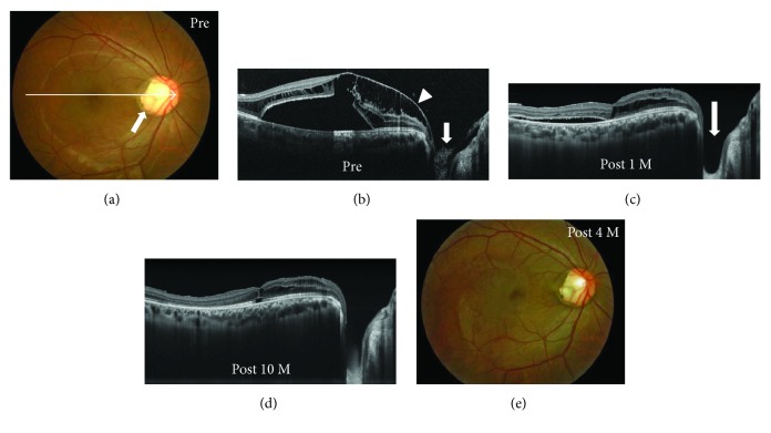
Figure 2.
Photographs of the right eye in a 24-year-old optic disc pit (ODP) maculopathy patient without posterior vitreous detachment (case 2). (a) Fundus photograph before surgery showed an ODP (arrow) and macular retinal elevation. An arrow indicates enhanced depth imaging optical coherence tomography (EDI-OCT) scans for the images shown in Figure 2. (b–d) EDI-OCT images. Before surgery, retinal schisis extended from the optic disc to the macula with macular detachment (b). Glial tissue suggests that the Cloquet's canal (b, arrow) was also present, invaginating into the ODP and the hyaloid membrane attached to the glial tissue (b, arrowhead). The retinal schisis was reduced 1 month after pars plana vitrectomy (c) and was almost resolved with foveal attachment 10 months after surgery (d). The epipapillary membrane tissue was also removed (c, arrow). (e) The retinal elevation was reduced at four months after surgery.

