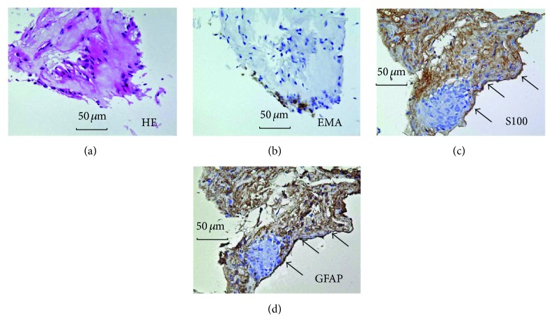
Figure 8.
Histology (a) and immunohistochemistry (b–d) of a resected epipapillary membrane tissue in case 5. (a) Light micrograph showing marked hyalinization. Spindle-shaped stromal cells were intermingled in the section (hematoxylin-eosin). (b) Immunochemistry for cytokeratin. Immunoreactivity was not observed. (c) Immunoreactivity for S100 was strongly detected in the membrane (arrows). (d) Immunoreactivity for GFAP was strongly detected in the membrane (arrows).

