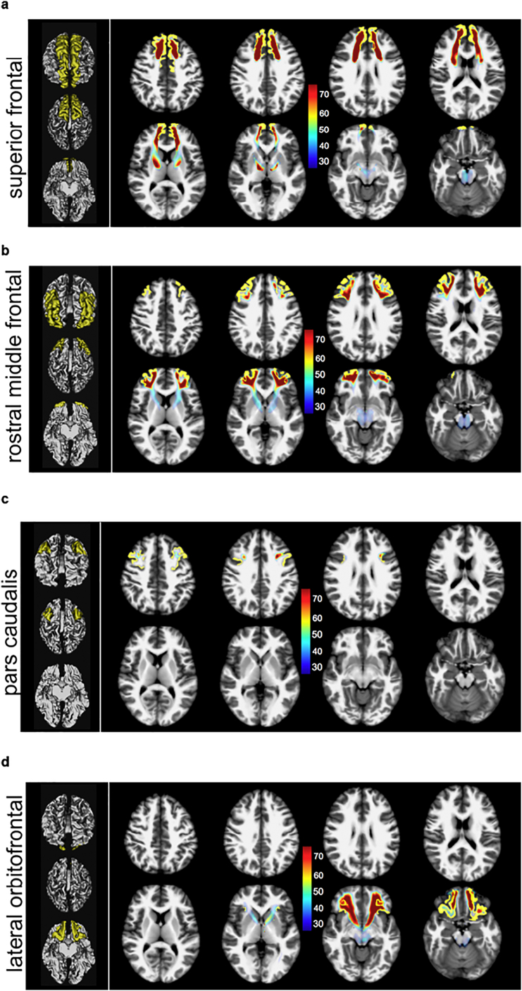
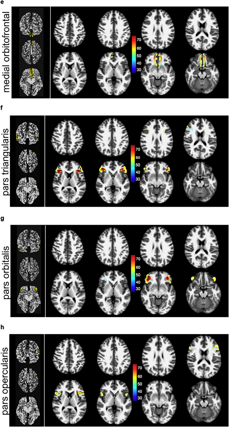
Fig. 5.
(a–h): slMFB and its prefrontal white matter (WM) sub-segments addressing distinct cortical parcellations of the PFC parceling according to Desikan/Killiany (Desikan et al., 2006) (axial slices, MNI space). Color scale shows probability of fiber occurrence in [%] relative to entire MFB structure. Global tractographic approach used. Due to the dominating nature of the superior frontal, rostral middle and lateral orbitofrontal segmentations, in these the trunk region of MFB shows up with roughly 30%.


