FIG 5.
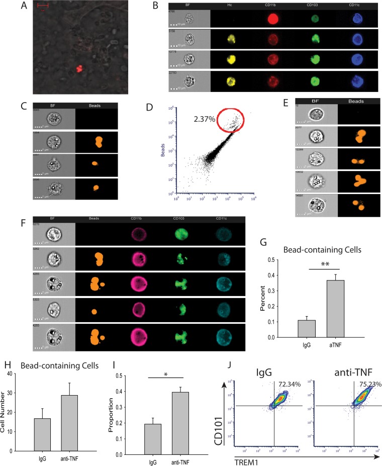
CD11b+ CD103+ DCs migrate from the gut to the lung. Mice were infected with 2 × 10⁶ H. capsulatum yeasts i.n. and treated with anti-TNF or isotype control antibody. (A and B) At the time of infection, 4 × 10⁶ tdTomato-H. capsulatum yeasts were delivered via oral gavage. Lungs were harvested at day 3 p.i. and assessed by confocal microscopy (A) and Amnis ImagestreamX (B). The data are representative of the results of two experiments containing 8 mice/group. BF, bright field. (C and D) Mice were treated with 4 × 10⁶ beads i.n., and lungs were harvested 3 days after treatment. Bead-containing cells were assessed by Amnis ImagestreamX. The data are representative of two mice. (E to H) Mice were treated with 4 × 10⁷ beads via oral gavage on days 0, 1, and 2 p.i. Lungs were harvested at day 3 p.i. and assessed by Amnis ImagestreamX. (I) Blood was drawn from the mice at day 2 p.i., and DCs were assessed by FACS. (J) Lungs were harvested at day 3 p.i., and DCs were assessed by FACS. The data represent means and SEM (n = 8 mice/group; representative of the results of two experiments). *, P < 0.05; **, P < 0.01.

