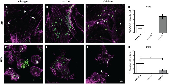
FIG 4.
Actin polymerization of wild-type R. parkeri compared to that of R. parkeri sca2::tn and rickA::tn in Vero and ISE6 cells at 48 hpi. Rickettsia (green) actively polymerizing actin (magenta) in Vero (A to C) and ISE6 (E to G) cells is shown. Wild-type R. parkeri (A and E), R. parkeri sca2::tn (B and F), and R. parkeri rickA::tn (C and G) strains are depicted. (D and H) Graphical representation of percent Rickettsia with actin tails in Vero (D) and ISE6 (H) cells. Data are representative of two replicates per experiment and two independent experiments. Statistical analysis consisted of a t test, with a P value of <0.05 being significant. White scale bar, 2 μm. Arrows indicate Rickettsia polymerizing actin.

