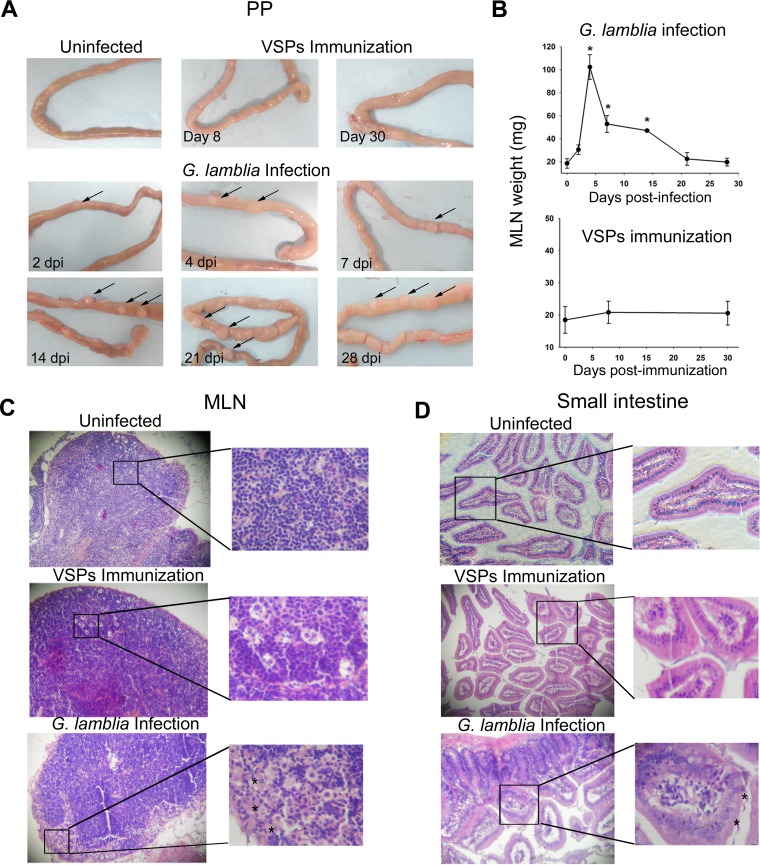
FIG 2.
Histopathological analysis. (A) Macroscopic examination of upper small intestines. Arrows indicate an increase in size of the Peyer's patches in the infected versus uninfected gerbils used as controls. (B) Mesenteric lymph node size. The MLN size from experimental gerbils was determined by weighing at the indicated day postinfection. Each point represents the mean value ± standard error of the mean of the results obtained in three independent experiments (n = 12). *, P < 0.05 compared to results in uninfected animals (0 dpi). (C and D) Light microscopy images. Representative images (magnification, ×20) from MLN and upper small intestine from uninfected, VSP-immunized, or infected gerbils are shown. Slides were stained by hematoxylin-eosin. Asterisks indicate some Giardia trophozoites in the intestinal lumen from infected gerbils (7 dpi) and eosinophilic areas enriched with histiocytes in an MLN section (4 dpi).

