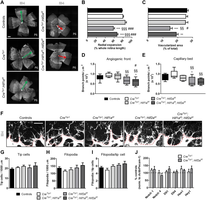
Fig. 3.
Deletion of Hif2a causes delayed developmental angiogenesis. (A-E) Isolectin B4 (iB4) staining of whole retinal flatmounts (A; age: P6) shows a delayed vascular development in Hif2a KO lines in terms of radial expansion (B), vascularized area (C), and number of branch points in the angiogenic front (D) and in the mature capillary bed (E). Green and red arrows indicate timely and delayed radial vascular expansion, respectively. n=5-39. Also refer to Fig. S2A. In D and E, whiskers are minimum and maximum value; line inside the box is the median. ANOVA and Tukey's multiple comparison test: *P<0.05, **P<0.01, ***P<0.001 vs controls; §§P<0.01, §§§P<0.001 vs CreTrp1; #P<0.05, ###P<0.001 vs CreTrp1;Hif1af/f. (F-I) Images (F) and quantification (G-I) of tip cells and filopodia at the angiogenic front (highlighted by the red dashed line) for all the genotypes analysed. n=6-19 (four fields of view/eye). No significant differences were observed (ANOVA). (J) Notch pathway gene expression assessed by RT-qPCR in FACS-sorted (iB4-FITC labelling) endothelial cells (age: P2). Results are expressed as percentage of controls (normalized to the endothelial marker Pecam1 and the housekeeping gene Actb); no significant differences (ANOVA). Each sample was a pool of 4-8 retinae; n=3-9. Scale bars: 500 µm (A); 25 µm (F).

